Understand Surgery: Femoral Popliteal Bypass

What is a Femoral Popliteal Bypass?
According to Stanford Health Care , a "femoral popliteal bypass is the surgical opening of the upper leg to directly visualize the femoral artery. It is performed to bypass the blocked portion of the artery using a piece of another blood vessel."
A-traumatic causes of AVN include alcohol consumption and the long term use of corticosteroids1, which may lead to peripheral vascular or peripheral arterial disease.2
A Two Minute Patient Education Video
Who needs a femoral popliteal bypass?
Patients who are diagnosed with the condition Avascular Necrosis (AVN),also known as osteonecrosis, may become candidates for the femoral popliteal bypass procedure. AVN means that the patient's bones are not receiving an adequate supply of blood, which may lead to chronic limb ischemia 4. Consequentially, without treatment, the bone marrow cells to start to die, and may even lead the affected bone to collapse.1 The most common cause of AVN is trauma, and usually occurs within 8 hours of receiving the injury. To clarify, this article focuses atraumatic arterial disease, diagnostics, and treatment.
Take a Guess!
view quiz statistics
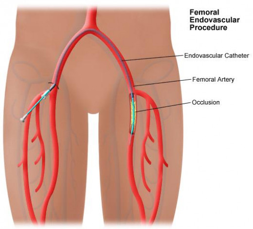
What are arteries?
Arteries are lumens, meaning that they have a tubular structure. They are made mostly out of smooth muscle and have thick protective outer walls. One of the most important characteristics of an artery is it's high elasticity, which allows the artery to contract during the heart phase systole, the action responsible for pumping blood throughout the body. All arteries move blood away from the heart to oxygenate and deliver nutrients to all body tissues. (Actually the pulmonary arteries have a different duty, to carry de-oxyginated blood to the lungs).5
Diagnostic Procedures
When an atherosclerosis occurs, fatty deposits and plaque build up have taken up residence somewhere in the patient's arteries. This is the primary cause of reduced blood flow within the artery. The arteries may become hardened, losing their elasticity, or ability to pump blood to tissue. In order to make a proper diagnosis, the medical team has to diagnose and identify the exact location of the occluded blood vessel.5
Diagnostic Procedure
| Description
|
|---|---|
Arterial Plethysmography
| Three blood pressure cuffs are placed on the patient's leg and inflated to 65 mm/hg. Each cuff produces a waveform which measures blood flow. A reduced wave-form may indicate reduced blood flow in that particular region.
|
Doppler Scanning
| Doppler scanning uses ultrasonic energy to identify abnormalities in blood flow, strictures, and thrombi turbulence ( the swirling motion of blood).
|
Arteriography
| Radiographic imaging is taken of an artery intraoperatively, or during a live interventional procedure to fix the shape and interior surface of an artery.
|
Intravascular Ultrasonography
| A catheter is inserted into the artery lumen and messure the density and accumulation of atherosclerotic plaque and wall thickness. It is capable of 3D rotation.
|
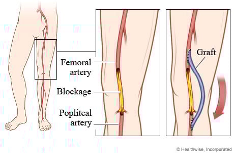
Imagine a soda straw chewed up and clogged in the center, completely unusable. Now cut the broken piece of straw out of the body of the straw. In order to make the straw functional again, measure a piece of a new straw and replace the chewed up part of the straw by joining the healthy ends of your original straw to your straw graft, that's what the goal of a femoralpopliteal bypass is, to circumvent the diseased, unusable artery and restore blood flow.

Surgical Treatment
After a femoral artery has been diagnosed with atherosclerosis surgical intervention may be ordered for the patient in one of two ways. If the plaque build up can be removed from the artery surgically, most likely the Surgeon will order an endartectomy. It is less invasive than a complete artery bypass, but it's not always a viable option depending on the integrity of the diseased artery itself.
The next surgical option is the femoralpopliteal bypass. The surgical goal of this procedure is to take either an autograft (removed from the patient) of the saphrenous vein, or a synthetic graft, and implant it between the femoral and popliteal arteries to bypass the diseased portion of the artery.
Fem-Pop Bypass in 11 Steps 5
1. An incision is made into the side of the thigh, down to the deep tissue.
2. The femoral artery is lifted and mobilized so that vessel loops can be placed around it.
3. An incision is made on the side of the knee.
4. The popliteal artery is identified and mobilized.
5. Angiograms are taken.
6. The graft is tunneled through the subcutatenous tissue and then connected to the two wound sites.
7. The femoral artery is clamped for control, ad a cut is made into the artery wall (arteriotomy), creating an opening.
8. One end (the proximal end) of the graft is attached lumen to lumen (anastomosed) to the femoral artery.
9. The popliteal anastomosis is performed at the other end of the graft.
10 Angiograms are performed to confirm the success of the operation by measuring pulse volume.
11.The wounds are closed.
Have you or a loved one suffered from peripheral vascular disease?
A 5-Star Guide to Living with Heart Disease

Citations
1. Taken from the Association of Surgical Technologists at AST.org
2. Taken from Stanford Healthcare at Stanfordhealthcare.org
3. Taken from the Health Library at Hopkinsmedicine.org
4. Taken from the US National Library for Medicine at ncbi.nlm.nih.gov
5. Joanna Kotcher Fuller, BA, BSN, RN, RGN, MPH (2013). Surgical Technology Principles and Practice, Sixth Edition. St. Louis, Missouri: Elsevier Inc.


